Imaging Center
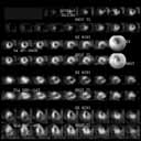
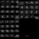
The Cardiac Imaging Center offers 
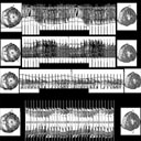
mutlilevel cardiac imaging including Nuclear Perfusion and functional 
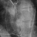
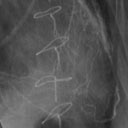
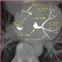
scans, Angiography, Structural, functional, coronary, and perfusion Magnetic 
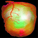
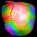
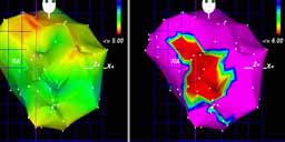
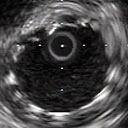
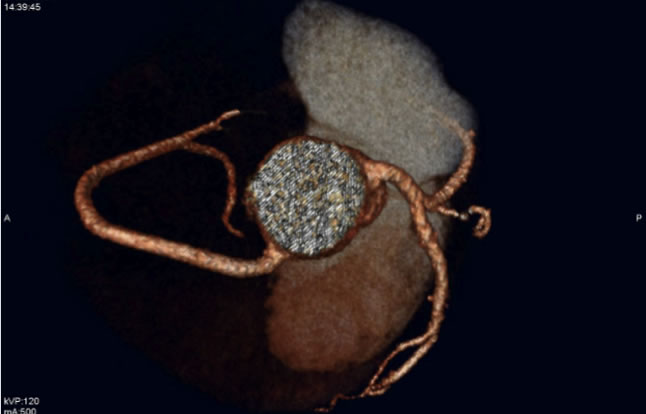
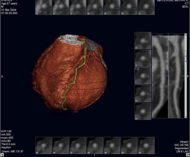
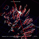
Resonance Imaging, 320 Detector Computerized Tomography, 2D, #D, and 4D Echocardiography, electromagnetic mapping, and 
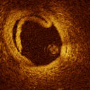
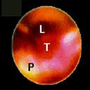
intravascular and Intracardiac Imaging, Optical Coherence Tomography, and Angioscopy. The Center functions as a core laboratory for several clinical trials of Angiogenesis as well as preclinical studies of angiogenesis, restenosis, and myocardial protection.

Functional Cardiac assessment Presentation:

Nuclear Perfusion and Functional Imaging: Left:Rest Thallium-Exercise MIBI shows inferior, anterior, and anterolateral ischemia Right: FDG SPECT showing viability in myocardial territories

PET Scanning with angiography and Biosense NOGA map overlay: Complex Image processing and image overlay allows researchers at the center to accurately and reproducibly evaluate various novel treatment options and target therapeutic agents to where they are needed

Transthoracic echocardiography (left) and transesophageal echocardiography (right): The availability of echocardiography and on and offline analysis allows longitudinal assessment of various treatment strategies in addition to accurate diagnosis in several clinical situations
Coronary Angiography and Digital Substraction Angiography: The center utilizes coronary angiographyin its preclinical and clinical studies to assess angiogenesis and restenosis. Shown is the RCA filling by Collaterals from the bypassed LAD in the heparin alginate FGF-2 study
Biosense NOGA electromechanical mapping: The Cardiac imaging center has extensive experience with Biosense mapping to diagnose ischemia, viability and to direct therapeutic intervention on the heart. An electromechanical map showing anterior dysfunction (red) with preserved voltage
Magnetic resonance imaging: The imaging center carries out MR imaging of the coronary arteries, ventricular structures, regional wall motion, perfusion, and collateralization both for clinical and preclinical use. Shown are 4D cardiac imaging and cine functional imaging
Intravascular imaging: This is performed using intravascular ultrasound (left_, optical coherence tomography (right) and intrvascular MRI. Functional imaging is done with pressure wires and Doppler flow wires to assess coronary blood flow
Electron Beam tomography (left): EBCT is done to assess the coronary circulation and to assess coronary calcifications. All these imaging modalities are analyzed using advanced image processing techniques (motion vs time, right) for core lab evaluation
Cardiac Computerized Tomography:Cardiac CT is done to assess the coronary anatomy, Myocardial perfusion, Plaque morphology, and chamber anatomy. This is performed using the 320 detector scanner allowing cardiac imaging within one cardiac cycle (seconds) reducing motion and splicing artifact. The center is particpating in several studies of cardiac CT (Core 320...)
Several other imaging modalities are available: including local trajectory assessment (left) and intracoronary angioscopy (right). All these modalities are available for clinical and preclinical use.