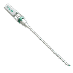
Cardiac catheterization
is a diagnostic procedure which is performed in order to detect problems
with the heart and its blood supply. A long, thin tube (a catheter) is
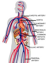
inserted into the femoral artery and vein to the heart and cardiac pressures
are measured and the catheters are used to inject X-Ray dye to visualize
the heart and the blood vessels. Newer catheter have allowed the use of
the wrist artery for certain patients (radial artery catheterization).
The catheterization procedure will provide a great deal of information
about the heart including the heart pumping ability (Ejection Fraction,
EF), the pressure within the various heart chambers, and detailed structural
information about the heart and blood vessels, and the pattern of flow
within the heart and major blood vessels.
The catheterization itself normally takes less than an hour and a simple heart catheterization is often an outpatient procedure. However, often, if a blockage is found that can be treated with balloon angioplasty, stenting, or other interventional techniques, the procedures will be performed in the same seting and the patient will stay overnight.
The catheterization itself normally takes less than an hour and a simple heart catheterization is often an outpatient procedure. However, often, if a blockage is found that can be treated with balloon angioplasty, stenting, or other interventional techniques, the procedures will be performed in the same seting and the patient will stay overnight.